Introduction to BiologyCorner.com’s Animal Cell Coloring Resource

Biologycorner.com animal cell coloring – BiologyCorner.com offers a valuable animal cell coloring resource designed to enhance learning and understanding of this fundamental biological concept. Coloring, often underestimated as a learning tool, provides a surprisingly effective way to engage students and solidify their knowledge of cell structures and functions. The interactive nature of coloring helps students actively participate in the learning process, making it more memorable and less passive than simply reading or listening to a lecture.Visual learning plays a crucial role in biology education, especially when dealing with complex structures like cells.
Our brains process visual information far more efficiently than text alone. By coloring the different organelles of an animal cell, students create a mental map of their locations and functions, strengthening their understanding and retention. This method caters to various learning styles, making it particularly beneficial for visual and kinesthetic learners.
Content and Features of BiologyCorner.com’s Animal Cell Coloring Resource
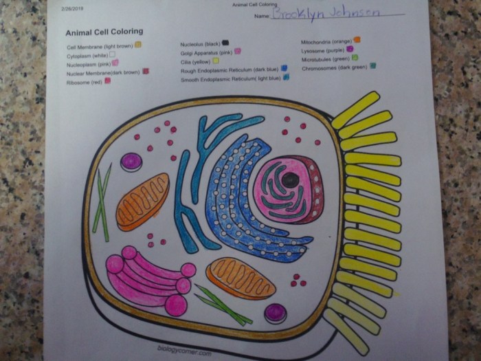
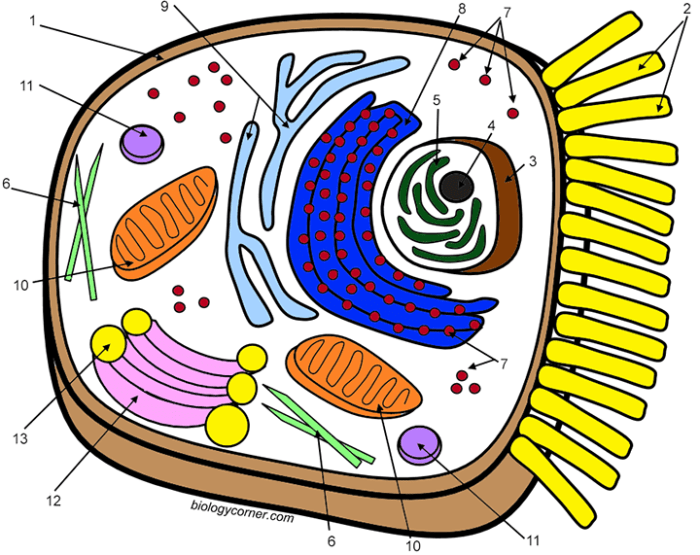
BiologyCorner.com’s animal cell coloring resource provides a printable worksheet featuring a detailed Artikel of an animal cell. The worksheet clearly labels key organelles such as the nucleus, mitochondria, ribosomes, endoplasmic reticulum, Golgi apparatus, lysosomes, and cell membrane. Students are encouraged to color each organelle using a specific color, further enhancing their memorization and association of structure with function.
This allows for a personalized learning experience, letting students customize their diagrams and add notes for extra reinforcement. The simple, clear design ensures the focus remains on the cell’s structure and avoids overwhelming students with excessive detail. Furthermore, the resource is readily available for download and printing, making it easily accessible for both individual and classroom use.
The detailed diagrams available at biologycorner.com for animal cell coloring provide a valuable educational resource, particularly for understanding cellular structures. For those seeking a broader range of illustrative activities, supplementing this with readily available resources like free animal coloring sheets can enhance learning and engagement. Returning to the intricacies of the animal cell, biologycorner.com offers a more focused approach to understanding cell biology through coloring.
The design is also compatible with various printing options, ensuring usability regardless of printer type or settings.
Animal Cell Structure and Function as Depicted in the Coloring Page
The animal cell coloring page provides a visual representation of the intricate machinery within a typical animal cell. Understanding the structure and function of each organelle is key to grasping how the cell operates as a whole, carrying out the processes necessary for life. This section will detail the key organelles and their roles as illustrated on the coloring page.The coloring page likely depicts several key organelles, each with a specific function crucial for cell survival.
The interactions between these organelles highlight the coordinated nature of cellular processes. We’ll explore these interactions and the importance of each component.
Cell Membrane
The cell membrane, often depicted as a thin outer boundary, is the selectively permeable barrier controlling what enters and exits the cell. This membrane is composed of a phospholipid bilayer, with embedded proteins facilitating transport. It maintains the cell’s internal environment, regulating the flow of nutrients, waste products, and signaling molecules. Without a functioning cell membrane, the cell cannot maintain homeostasis and will not survive.
Cytoplasm
The cytoplasm, the jelly-like substance filling the cell, is the site of many metabolic reactions. It’s a dynamic environment where organelles are suspended and where many crucial biochemical processes occur. The cytoplasm provides a medium for the transport of molecules and facilitates cellular communication. Its consistency and composition are essential for maintaining cellular structure and function.
Nucleus
The nucleus, often shown as a large, centrally located structure, is the control center of the cell. It houses the cell’s genetic material (DNA), which directs all cellular activities. The nucleus regulates gene expression, controlling which proteins are synthesized and when. The nuclear membrane, a double membrane surrounding the nucleus, controls the passage of molecules between the nucleus and cytoplasm.
Damage to the nucleus severely compromises the cell’s ability to function and reproduce.
Ribosomes
Ribosomes, often depicted as small dots scattered throughout the cytoplasm and on the endoplasmic reticulum, are the protein synthesis factories of the cell. They translate the genetic code from mRNA into proteins, the workhorses of the cell. Ribosomes are essential for building all the proteins needed for cell structure, function, and regulation. A deficiency in ribosome function would severely limit a cell’s ability to produce necessary proteins.
Endoplasmic Reticulum (ER)
The endoplasmic reticulum (ER), often shown as a network of interconnected membranes, comes in two forms: rough ER (studded with ribosomes) and smooth ER (lacking ribosomes). Rough ER is involved in protein synthesis and modification, while smooth ER synthesizes lipids and detoxifies certain substances. The ER’s extensive network allows for efficient transport of molecules within the cell. Its absence would severely disrupt protein synthesis and lipid metabolism.
Golgi Apparatus
The Golgi apparatus, often depicted as a stack of flattened sacs, is the cell’s processing and packaging center. It receives proteins and lipids from the ER, modifies them, and sorts them for transport to their final destinations within or outside the cell. The Golgi apparatus is crucial for proper protein function and secretion. Its dysfunction would lead to the accumulation of improperly processed molecules.
Mitochondria
Mitochondria, often depicted as bean-shaped organelles, are the powerhouses of the cell. They generate ATP (adenosine triphosphate), the cell’s main energy currency, through cellular respiration. Mitochondria are essential for providing energy for all cellular processes. Mitochondrial dysfunction is linked to numerous diseases due to energy deficits within the cells.
Lysosomes
Lysosomes, often depicted as small, membrane-bound sacs, contain digestive enzymes that break down waste products, cellular debris, and foreign materials. They are crucial for maintaining cellular cleanliness and preventing the buildup of harmful substances. Lysosomal dysfunction can lead to the accumulation of undigested materials within the cell, causing damage.
Creating an Enhanced Coloring Page with Added Details
A simple coloring page can be a great starting point for learning about animal cell structure, but adding detailed labels significantly enhances the educational value. By incorporating more specific labels and strategically placing them, we can create a more effective learning tool that improves comprehension and retention of key biological concepts. This improved design moves beyond simple identification to a deeper understanding of organelle function.The modified BiologyCorner.com animal cell coloring page would feature a more detailed representation of the cell’s components.
Each organelle would not only be visually distinct but also clearly labeled with its name. Furthermore, the arrangement of labels would be carefully considered to prevent visual clutter and ensure easy reading. The goal is to create a visually appealing and pedagogically sound resource.
Label Placement and Organization on the Enhanced Coloring Page
The placement of labels is crucial for clarity. We would avoid overlapping labels and ensure sufficient space between them and the organelles they identify. A consistent font size and style would maintain visual uniformity. For example, the nucleus would be clearly labeled with “Nucleus” directly adjacent to its illustrated representation, but not obscuring the drawing of the nuclear membrane or nucleolus.
Similarly, smaller organelles like ribosomes might be grouped together with a collective label and a small arrow pointing to a cluster of them. This method prevents overwhelming the image with too many individual labels while still providing sufficient detail. Labels could be color-coded to match the organelle’s color in the drawing for enhanced visual association. For instance, the rough endoplasmic reticulum, often depicted in a blueish-gray, could have its label in the same shade.
Improved Learning Experience through Enhanced Labeling
The addition of detailed labels transforms the coloring page from a passive activity to an active learning experience. The process of coloring and labeling simultaneously reinforces visual memory and strengthens conceptual understanding. Students actively engage with the material, associating the visual representation of each organelle with its specific name and function. This active engagement leads to better knowledge retention compared to simply reading a textbook description.
For example, a student coloring the mitochondria and simultaneously labeling it will have a much stronger association between the organelle’s visual representation and its function in cellular respiration than if they only read about mitochondria. The enhanced coloring page provides a multi-sensory learning experience, improving the overall effectiveness of the educational material.
Illustrative Description of Cellular Processes

Let’s delve into the fascinating world of cellular processes, specifically protein synthesis and cellular respiration, and see how they’re visually represented in an animal cell coloring page. Understanding these processes highlights the crucial interplay between a cell’s structure and its function.Protein synthesis, the creation of proteins, is a multi-step process involving several organelles. The coloring page can visually represent this by showing the nucleus (containing DNA), the ribosomes (where protein synthesis occurs), the endoplasmic reticulum (ER) (modifying and transporting proteins), and the Golgi apparatus (packaging and shipping proteins).
The different stages of protein synthesis – transcription (DNA to mRNA) and translation (mRNA to protein) – could be conceptually illustrated through color-coding or distinct shapes representing different molecules involved.
Protein Synthesis: A Detailed Look
The nucleus, depicted as a large, centrally located circle in the coloring page, houses the cell’s DNA, the blueprint for protein creation. Transcription, the process of creating messenger RNA (mRNA) from DNA, happens inside the nucleus. This mRNA then travels to the ribosomes, which can be shown as small dots scattered throughout the cytoplasm or attached to the endoplasmic reticulum (ER).
Ribosomes are the sites of translation, where the mRNA code is read, and amino acids are assembled into a polypeptide chain, forming the protein. The rough endoplasmic reticulum (RER), illustrated as a network of interconnected membranes studded with ribosomes in the coloring page, modifies and folds the newly synthesized proteins. The Golgi apparatus, shown as a stack of flattened sacs, further processes, sorts, and packages the proteins into vesicles for transport to their final destinations within or outside the cell.
The coloring page could visually distinguish the RER from the smooth ER (lacking ribosomes) through different shading or coloring. This detailed representation helps students visualize the coordinated actions of multiple organelles.
Cellular Respiration: Energy Production
Cellular respiration, the process of generating energy (ATP) from glucose, is another vital cellular process. The coloring page can illustrate this by highlighting the mitochondria, the powerhouse of the cell, depicted as bean-shaped organelles with internal cristae (folds). The process begins in the cytoplasm with glycolysis, breaking down glucose into pyruvate. The pyruvate then enters the mitochondria, where the Krebs cycle (citric acid cycle) and oxidative phosphorylation occur.
These stages can be visually differentiated within the mitochondria’s illustration on the coloring page, perhaps through different color-coding of the matrix and cristae. The production of ATP, the cell’s energy currency, could be symbolized by a specific color or pattern within the mitochondria. The smooth endoplasmic reticulum, often depicted as a network of interconnected membranes without ribosomes, plays a role in lipid metabolism, which provides fuel for cellular respiration.
The coloring page might subtly distinguish it from the rough ER through differences in texture or color.
Structure-Function Relationship in Cellular Processes
The intricate structure of the animal cell is directly linked to its ability to carry out complex processes like protein synthesis and cellular respiration. For instance, the compartmentalization provided by organelles such as the nucleus, ER, and Golgi apparatus is essential for efficient protein synthesis, preventing the mixing of different steps and molecules. Similarly, the highly folded inner membrane of the mitochondria (cristae) significantly increases the surface area available for the electron transport chain, maximizing ATP production during cellular respiration.
The coloring page can effectively highlight this structure-function relationship by clearly depicting the unique morphology of each organelle and its position within the cell, showcasing how their spatial arrangement facilitates the smooth execution of these essential processes. For example, the proximity of ribosomes to the ER efficiently channels newly synthesized proteins for modification.
Extension Activities Based on the Coloring Page: Biologycorner.com Animal Cell Coloring

The animal cell coloring page serves as a fantastic foundation for deeper learning. These activities are designed to move beyond simple identification and encourage a more active and engaged understanding of animal cell structure and function. They’re structured to cater to various learning styles and levels of prior knowledge.
The following extension activities offer diverse approaches to solidify understanding and promote further exploration of animal cell biology. They range from creative projects to more analytical investigations, all designed to reinforce learning in a dynamic and engaging manner.
Animal Cell Analogies and Models
Creating analogies helps students connect abstract cellular concepts to familiar objects. For example, students could compare the cell membrane to a castle wall, protecting the cell’s contents, while the nucleus could be likened to the castle’s control center. They could build 3D models of animal cells using readily available materials like balloons, pipe cleaners, and colored candies, assigning each material to a specific organelle and explaining its function.
This hands-on approach reinforces understanding of organelle structure and their relative sizes within the cell.
Researching Specific Organelles
Students can choose an organelle from the coloring page (e.g., mitochondria, Golgi apparatus, ribosomes) and research its specific function in more detail. They could then present their findings in a short report, poster, or presentation, including diagrams and visual aids to enhance understanding. For example, research on mitochondria could explore its role in cellular respiration and energy production, highlighting the process of ATP synthesis.
Research on the Golgi apparatus could focus on its role in protein modification and packaging for secretion.
Cell Processes Diagrams and Explanations
Students can create detailed diagrams illustrating key cellular processes such as protein synthesis (transcription and translation), cellular respiration, or cell division (mitosis). These diagrams should not only depict the process visually but also include written explanations detailing each step and the involvement of specific organelles. For example, a diagram of protein synthesis could illustrate the movement of mRNA from the nucleus to the ribosomes in the cytoplasm, the role of tRNA in bringing amino acids, and the formation of a polypeptide chain.
Similarly, a diagram illustrating cellular respiration could show the breakdown of glucose in the mitochondria and the production of ATP.
Comparative Cell Biology, Biologycorner.com animal cell coloring
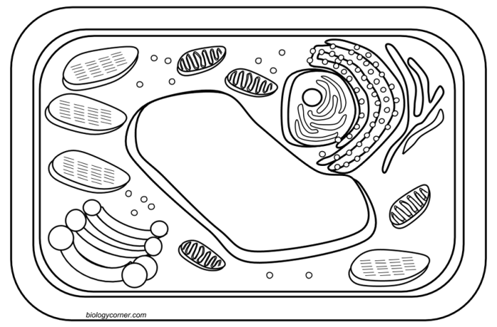
Students can compare and contrast animal cells with other types of cells, such as plant cells or bacterial cells. This comparison could focus on the presence or absence of specific organelles, the differences in cell wall structure (if present), and the overall size and shape of the cells. For instance, a comparison could highlight the presence of a cell wall and chloroplasts in plant cells, absent in animal cells, and discuss the implications of these differences in terms of cellular function and overall organismal characteristics.
FAQ
Is this coloring page suitable for all ages?
Yes, it can be adapted for various age groups. Younger children can focus on basic identification, while older students can delve into more complex functions and processes.
Can I print the coloring page directly from the website?
Yes, Biologycorner.com provides a printable version of the animal cell coloring page.
Are there any answer keys available?
While a formal answer key might not be explicitly provided, the website’s content offers sufficient information to identify the organelles.
What other resources does Biologycorner.com offer?
Biologycorner.com offers a wide range of educational resources beyond this coloring page, including other worksheets, quizzes, and interactive lessons.

